Wednesday, March 16, 2011

Clubfoot


Parents know immediately if their newborn has a clubfoot. Some will even know before the child is born, if an ultrasound was done during the pregnancy. A clubfoot occurs in approximately one in every 1,000 births, with boys slightly outnumbering girls. One or both feet may be affected.


Cause

Doctors still aren't certain why it happens, though it can occur in some families with previous clubfeet. In fact, your baby's chance of having a clubfoot is twice as likely if you, your spouse or your other children also have it. Less severe infant foot problems are common and are often incorrectly called clubfoot.

Symptoms


The appearance is unmistakable: the foot is turned to the side and it may even appear that the top of the foot is where the bottom should be.

The involved foot, calf, and leg are smaller and shorter than the normal side.

It is not a painful condition. But if it is not treated, clubfoot will lead to significant discomfort and disability by the teenage years.

Treatment


Nonsurgical Treatment

Treatment should begin right away to have the best chance for a successful outcome without the need for surgery. Over the past 10 to 15 years, more and more success has been achieved in correcting clubfeet without the need for surgery. A particular method of stretching and casting, known as the Ponseti method, has been responsible for this. With this method, the doctor
changes the cast every week for several weeks, always stretching the foot toward the correct position. The heel cord is then released followed by one more cast for three weeks.

Once the foot has been corrected, the infant must wear a brace at night for two years to maintain the correction. This has been extremely effective but requires the parents to actively participate in the daily care by applying the braces. Without the parents' participation, the clubfoot will almost certainly recur. That's because the muscles around the foot can pull it back into the abnormal position.

The goal of this, and any treatment program, is to make your newborn's clubfoot (or feet) functional, painless and stable by the time he or she is ready to walk. (Note: Anytime your baby wears a cast, watch for changes in skin color or temperature that may indicate problems with circulation.)

Surgical Treatment

On occasion, stretching, casting and bracing are not enough to correct your baby's clubfoot. Surgery may be needed to adjust the tendons, ligaments and joints in the foot/ankle. Usually done at 9 to12 months of age, surgery corrects all of your baby's clubfoot deformities at the same time. After surgery, a cast holds the clubfoot still while it heals. It's still possible for the muscles in your child's foot to try to return to the clubfoot position, and special shoes or braces will likely be used for up to a year or more after surgery. Surgery will likely result in a stiffer foot than nonsurgical treatment, particularly as the years pass by.

Without any treatment, your child's clubfoot will result in severe functional disability. With treatment, your child should have a nearly normal foot. He or she can run and play without pain and wear normal shoes. The corrected clubfoot will still not be perfect, however. You should expect it to stay 1 to 1 1/2 sizes smaller and somewhat less mobile than the normal foot. The calf muscles in your child's clubfoot leg will also stay smaller.

Claw Toe


People often blame the common foot deformity claw toe on wearing shoes that squeeze your toes, such as shoes that are too short or high heels. However, claw toe also is often the result of nerve damage caused by diseases like diabetes or alcoholism, which can weaken the muscles in your foot. Having claw toe means your toes "claw," digging down into the soles of your shoes
and creating painful calluses. Claw toe gets worse without treatment and may become a permanent deformity over time.


Symptoms

• Your toes are bent upward (extension) from the joints at the ball of the foot.
• Your toes are bent downward (flexion) at the middle joints toward the sole of your shoe.
• Sometimes your toes also bend downward at the top joints, curling under the foot.
• Corns may develop over the top of the toe or under the ball of the foot.


Evaluation

If you have symptoms of a claw toe, see your doctor for evaluation. You may need certain tests to rule out neurological disorders that can weaken your foot muscles, creating imbalances that bend your toes. Trauma and inflammation can also cause claw toe deformity.

Treatment

Claw toe deformities are usually flexible at first, but they harden into place over time. If you have claw toe in early stages, your doctor may recommend a splint or tape to hold your toes in correct position. Additional advice:

• Wear shoes with soft, roomy toe boxes and avoid tight shoes and high-heels.
• Use your hands to stretch your toes and toe joints toward their normal positions.
• Exercise your toes by using them to pick up marbles or crumple a towel laid flat on the floor.

If you have claw toe in later stages and your toes are fixed in position:

• A special pad can redistribute your weight and relieve pressure on the ball of your foot.
• Try special "in depth" shoes that have an extra 3/8" depth in the toe box.
• Ask a shoe repair shop to stretch a small pocket in the toe box to accommodate the deformity.

If these treatments do not help, you may need surgery to correct the problem.

Chordoma


Chordoma is a rare tumor that usually occurs in the spine and base of the skull. It is a malignant tumor that grows fairly slowly. It can spread to other organs, usually the lungs. It represents only about 1 percent of all malignant bone tumors.

Description

Chordomas develop in a structure called the notochord. The notochord forms the early spine in the beginning stages of fetal development. Most of the notochord is replaced by the spine during the first six months of development. Small areas can remain, and chordomas can form in these areas.

Most chordomas occur at the base of the spine (sacrum), in the tailbone (coccyx) or at the base of the skull (40 percent), but they can occur other places in the spine.

Most patients with chordomas are between 40 and 70 years of age. Occasionally this tumor can occur in younger patients, even children. The average age of people affected is about 55 years.

Chordomas are life-threatening. They can cause death by direct growth or by spreading to other organs. They spread to the lungs about 20 to 30 percent of the time.

Cause

Chordomas occur spontaneously. They are not known to be caused by trauma, environmental factors, or diet. Chordomas are not inherited. Chordomas are not associated with any medical conditions or use of any medications or supplements.

Symptoms

Chordomas grow slowly. Patients often have symptoms for a long time (more than a year) before they seek medical attention.

Symptoms depend on the location of the tumor:

• Tumors in the skull can cause headache or problems with vision.
• Tumors in the spine can cause pain in the area of tumor (neck, back, or tailbone). A tumor pressing on nearby nerves can cause symptoms similar to a "slipped" (herniated) disk. These symptoms include arm or leg pain, weakness, or numbness.
• Tumors in the tailbone area can cause a noticeable mass, numbness in the crotch area, and problems with the bowel and bladder.

Tests

Several imaging tests may be required to identify a chordoma, including x-rays, computed tomography (CT) scans, and magnetic resonance imaging (MRI) scans.

Chordomas themselves do not show up well, but the bone damage may be seen on the x-rays. X-rays of the sacrum are often difficult for doctors to interpret.


Typical sacral chordomas.


Computed tomography (CT) scans of the same sacral chordomas.


Corresponding magnetic resonance (MRI) scans.


Treatment

Treatment of chordomas is very difficult. This is mainly because these tumors are near the brain and spinal cord.

Traditional chemotherapy has not been very effective, and radiation alone rarely cures the tumor.

Surgery is the preferred treatment option, where possible. Surgery in combination with radiation is often used to treat chordomas. When tumors cannot be surgically removed, sometimes radiation therapy can stop the growth of the tumor.

Although it is the best treatment for chordomas, in many cases surgery can be difficult to perform because of important structures near the tumor. The surgeon must remove the tumor and a margin of normal tissue around it. This often results in a loss of neurologic function. For example, surgical removal of tumors in the sacrum can result in loss of control of bowel and bladder function (incontinence).

Fifty to 75% of patients treated with surgery will be alive after 5 years.

Research on the Horizon

Advances in radiation therapy are allowing higher doses of radiation to be applied with less damage to surrounding normal tissues, such as the brain and spinal cord. Different types of radiation therapy (including proton beam therapy, intraoperative radiation, and brachytherapy) may offer better treatment of these tumors. These therapies may be combined with more limited surgery in the future.

New drug treatments, including use of a drug called imatinib mseylate are being investigated which may help slow the growth of tumors that cannot be removed.

Cervical Spondylotic Myelopathy


Cervical Spondylotic Myelopathy (Spinal Cord Compression) 

One of the most common neck conditions that occurs with age is cervical spondylotic myelopathy (CSM). Over time, the normal wear-and-tear effects of aging can lead to a narrowing of the spinal canal. This compresses — or squeezes — the spinal cord. CSM can cause a variety of symptoms, including pain, numbness, and weakness.

Understanding your spine and how it works can help you better understand CSM.


Description 


The vertebral column.

Spinal cord compression is a very common neck condition affecting older people in the United States. Symptoms usually begin after the age of 50, but can occur earlier if there was an injury to the spine at a younger age.

Many people with CSM will have steady progression of their disease. Once symptoms start, they tend to continue. Typically, the disease progresses slowly over several years. In about 5% to 20% of people, CSM worsens more rapidly.

Cause

Neck pain may result from abnormalities in the soft tissues—the muscles, ligaments, and nerves—as well as in bones and joints of the spine. The most common causes of neck pain are soft-tissue abnormalities due to injury or prolonged wear and tear. In rare instances, infection or tumors may cause neck pain. In some people, neck problems may be the source of pain in the
upper back, shoulders, or arms.

Inflammatory Diseases

Rheumatoid arthritis can destroy joints in the neck and cause severe stiffness and pain. Rheumatoid arthritis typically occurs in the upper neck area.

Cervical Disk Degeneration 


The disk acts as a shock absorber between the bones in the neck. In cervical disk degeneration (which typically occurs in people age 40 years and older), the normal gelatin-like center of the disk degenerates and the space between the vertebrae narrows. As the disk space narrows, added stress is applied to the joints of the spine causing further wear and degenerative disease.

The cervical disk may also protrude and put pressure on the spinal cord or nerve roots when the rim of the disk weakens. This is known as a herniated cervical disk.


(Left) Side view of a healthy cervical vertebra and disk. (Right) A disk that has degenerated and collapsed.

Injury

Because the neck is so flexible and because it supports the head, it is extremely vulnerable to injury. Motor vehicle or diving accidents, contact sports, and falls may result in neck injury.

The regular use of safety belts in motor vehicles can help to prevent or minimize neck injury. A "rear end" automobile collision may result in hyperextension, a backward motion of the neck beyond normal limits, or hyperflexion, a forward motion of the neck beyond normal limits.

The most common neck injuries involve the soft tissues: the muscles and ligaments. Severe neck injuries with a fracture or dislocation of the neck may damage the spinal cord and cause paralysis.

Other Causes

Less common causes of neck pain include tumors, infections, or congenital abnormalities of the vertebrae.

Symptoms 

The spinal cord is the cable of nerves that send and receive signals from the body. When the spinal cord is slowly compressed, people may develop symptoms such as:

• Tingling and numbness
• Weakness: trouble lifting objects, dropping things
• Difficulty walking (loss of balance), wide-based gait
• Coordination problems/clumsiness: handwriting, buttoning clothes, and feeding oneself can become difficult
• Neck pain and stiffness

Doctor Examination

After discussing your symptoms and medical history, your doctor will examine your neck. He or she will look for:

• Abnormal reflexes: usually overactive (hyper-reflexia)
• Weakness: usually in the arms more than in the legs
• Numbness: mostly in the arms and hands
• Atrophy: muscles deteriorate and shrink


Tests 

This MRI image shows bulging disks pressing on the spinal cord.

Other tests which may help your doctor confirm your diagnosis include:

X-rays 

These studies are useful to look at the alignment of your neck.

Magnetic resonance imaging (MRI). 

These studies can create better images of soft tissues. They can be used to look for compression of the spinal cord.

Myelogram

Your doctor may order this special type of computed tomography (CT) scan. CT scans allow better detailed cross-section images showing bone spurs and the size of the spinal canal. In a myelogram, color dye is injected before the scan to better outline the spinal cord.


Treatment 

Nonsurgical Treatment 

Some people get relief from symptoms without surgery. The most common nonsurgical treatment options include:

Soft Collars. Soft collars allow the muscles of the neck to rest and limit neck motion. This can help decrease pinching of nerve roots with movement. Soft collars should only be worn for short periods of time, because long-term wear can decrease the strength of neck muscles.

Exercise. Improving neck strength and flexibility with simple exercises may lessen discomfort.

Nonsteroidal anti-inflammatory medications (NSAIDs). Drugs like aspirin and ibuprofen can reduce swelling and painful symptoms.

Epidural steroid injections. Cortisone is a powerful anti-inflammatory. Cortisone injections in the "epidural space" can decrease swelling as well as pain, although they are not often used in CSM.

Chiropractic manipulation. Manipulation is never used if a patient has spinal cord
compression.

Surgical Treatment

Whether to have surgery or not is a complex decision made between you and your doctor.

Cervical Spondylosis


Cervical Spondylosis (Arthritis of the Neck)


Neck pain is extremely common. It can be caused by many things, and is most often related to getting older.

Like the rest of the body, the bones in the neck (cervical spine) slowly degenerate as we age. This frequently results in arthritis. Arthritis of the neck is called cervical spondylosis.

Cervical spondylosis is the degeneration of the joints in the neck. It becomes increasingly more common as people age. More than 85% of people over age 60 are affected.

Although it is a form of arthritis, cervical spondylosis rarely becomes a crippling or disabling type.
Understanding your spine and how it works can help you better understand cervical spondylosis.


Cause


Disk Degeneration and Bone Spurs

In the spine, arthritis can result as the disk degenerates and loses water content. In children and young adults, disks have high water content. As we get older, our disks begin to dry out and weaken. This problem causes settling, or collapse, of the disk spaces and loss of disk space height.


As the facet joints experience increased pressure, they also begin to degenerate and develop arthritis, similar to the hip or knee joint. The cartilage that covers and protects the joints wears away.

If the cartilage wears away completely, it can result in bone rubbing on bone. To make up for the lost cartilage, your body may respond by growing new bone in your facet joints to help support the vertebrae. Over time, this bone overgrowth — called spurs — may narrow the space for the nerves to pass through (stenosis).

(Left) Side view of a healthy cervical vertebra and disk. (Right) A disk that has degenerated and collapsed.

Risk Factors

There are several factors that increase your risk for cervical spondylosis. The following have all been linked to higher risks of neck pain and spondylosis:

• Genetics -if your family has a history of neck pain
• Smoking -clearly linked to increased neck pain
• Occupation -jobs with lots of neck motion and overhead work
• Mental health issues -depression/anxiety
• Injuries/trauma -car wreck or on-the-job injury

Symptoms

Pain from cervical spondylosis can be mild to severe. It is sometimes worsened by looking up or down for a long time, or with activities such as driving or reading a book. It also feels better with rest or lying down.

Additional symptoms include:

• Neck pain and stiffness (may be worse with activity)
• Numbness and weakness in arms, hands, and fingers
• Trouble walking, loss of balance, or weakness in hands or legs
• Muscle spasms in neck and shoulders
• Headaches
• Grinding and popping sound/feeling in neck with movement


Doctor Examination


Determining the source of the pain is essential to recommend the appropriate treatment and rehabilitation. Therefore, a comprehensive examination is required to determine the cause of neck pain.

Your doctor will take a complete history of the difficulties you are having with your neck. He or she may ask you about other illnesses or injuries that occurred to your neck. Questions may include: When did your neck begin to hurt? Has it ever hurt like this before? When your neck hurts, how often and for how long does it hurt? Does anything make it better or worse? Were you ever involved in an accident or had an injury to your neck? Have you ever been treated for your
neck pain?

A thorough physical exam will include your neck, shoulders, arms, and frequently your legs, as well. Your strength, touch sensation, reflexes, blood flow, flexibility of your neck and arms as well as your walking may be tested. The doctor may press on your neck and shoulders, and feel for trigger (tender) points or swollen glands.

Tests 

Your doctor may supplement your evaluation with blood tests, and, if necessary, consult with other medical specialists. Other tests which may help your doctor confirm your diagnosis include:

X-rays

These pictures are traditionally ordered as a first step in imaging the spine. X-rays will show aging changes, like loss of disk height or bone spurs.

Magnetic resonance imaging (MRI)

This study can create better images of soft tissues, such as muscles, disks, nerves, and the spinal cord.

Computed tomography (CT) scans

This specialized x-ray study allows careful evaluation of the bone and spinal canal.

Myelography 

This specific x-ray study involves injecting dye or contrast material into the spinal canal. It allows for careful evaluation of the spinal canal and nerve roots.

Electromyography (EMG) 

Nerve conduction studies and electromyography may be performed by another doctor to look for nerve damage or pinching.

Treatment 

Nonsurgical Treatment 

Physical therapy. Strengthening and stretching weakened or strained muscles is usually the first treatment that is advised. Your physical therapist may also use cervical (neck) traction and posture therapy. Physical therapy programs vary, but they generally last from 6 to 8 weeks.
Sessions are scheduled 2 to 3 times a week.

Medications. Several medications may be used together during the first phase of treatment to address both pain and inflammation.

• Acetaminophen. Mild pain is often relieved with acetaminophen.
• Non-steroidal anti-inflammatory drugs (NSAIDs). Often prescribed with
acetaminophen, drugs like ibuprofen and and naproxen are considered first-line
medicines for neck pain. They address both pain and swelling, and may be prescribed for a number of weeks, depending on the specific problem. Other types of pain medicines can be considered if you have serious contraindications to NSAIDs, or your pain is not well controlled.
• Muscle relaxants. Medications such as cyclobenzaprine or carisoprodol can also be used in the case of painful muscle spasms.

Soft Collars. These collars limit neck motion and allow the muscles of the neck to rest. Soft collars should only be worn for short periods of time because long-term wear can decrease the strength of neck muscles.

Ice, heat, other modalities. Careful use of ice, heat, massage, and other local therapies can help relieve symptoms.

Steroid-Based Injections. Many patients find short-term pain relief from steroid injections. Various types of these injections are routinely performed. The most common procedures for neck pain include:

Cervical epidural block. In this procedure, steroid and anesthetic medicine is injected into the space next to the covering of the spinal cord ("epidural" space). This procedure is typically used for neck and/ or arm pain that may be due to a cervical disk herniation, also known as radiculopathy or a "pinched nerve."


Epidural injection in the cervical spine. 

Cervical facet joint block. Steroid and anesthetic medicine is injected into the capsule of the facet joint in this procedure. The facet joints are located in the back of the neck and provide stability and movement. These joints can develop arthritic changes that may contribute to neck pain.


Cervical facet joint. 

Medial branch block and radiofrequency ablation. This procedure is used in some cases of chronic neck pain. It can be used for both diagnosis and treatment of a potentially painful joint.


Facet joint injection in the cervical spine.

During the diagnosis portion of the procedure, the nerve that supplies the facet joint is blocked with a local anesthetic (like the medicine used by your dentist). Your doctor will ask if your neck pain is completely gone. If so, then your doctor has pin-pointed the source of your neck pain.
The next step is to block the pain more permanently. This is done by damaging the nerve with radiofrequency, a procedure called radiofrequency ablation.

Although less invasive than surgery, steroid-based injections are prescribed only after a complete evaluation by your doctor. Before considering these injections, discuss with your doctor the risks and benefits of these procedures for your specific condition.

Surgical Treatment 

It is uncommon for people with only cervical spondylosis and neck pain to be treated with surgery. Surgery is reserved for patients who have severe pain that has not been relieved by other treatment. Some patients with severe pain will unfortunately not be candidates for surgery. This is due to the widespread nature of their arthritis, other medical problems, or other causes for their
pain, such as fibromyalgia.


People who have progressive neurologic symptoms, such as weakness, numbness, or falling, are more likely to be helped by surgery.

Cervical Radiculopathy


Cervical Radiculopathy (Pinched Nerve) 


Some people have neck pain that may radiate into the shoulder and arm. This type of pain is often caused by an injury near the root of a spinal nerve. A nerve root injury is sometimes referred to as a "pinched" nerve. The medical term for this condition is cervical radiculopathy.

Understanding your spine and how it works can help you better understand cervical radiculopathy. Learn more about your spine: Spine Basics

Cause 

As disks age, they lose height and begin to bulge. They also lose water content and become stiffer.

(Left) Side view of a healthy cervical vertebra and disk. (Right) A disk that has degenerated and collapsed.

As the disks lose height, the vertebrae move closer together. The body sees the collapsed disk as a possible weak area and responds by forming more bone — called spurs — around the disk to strengthen it. The bone spurs that form also contribute to the stiffening of the spine. Bone spurs may also narrow the area of the foramen and pinch the nerve root.

The disk changes that occur with age are often called arthritis or spondylosis. It is important to keep in mind that all these changes are "normal" and they occur in everyone. In fact, if MRI scans were performed on all people aged 50 or older, nearly half of the scans would show worn disks and pinched nerves that do not cause painful symptoms. It is not known why some patients have symptoms and others do not.

Symptoms

Cervical radiculopathy pain travels down the arm in the area of the involved nerve. Pain is usually described as sharp. There can also be a "pins and needles" sensation or even complete numbness. In addition, there may be a feeling of weakness with certain actvities.

Symptoms can be worsened with certain movements, like extending or straining the neck or turning the head. These symptoms are often made better by placing the hand on the head and stretching the shoulder.

Doctor Examination 

After discussing your medical history and symptoms, your doctor will examine your neck. This will include testing your strength and sensation as well as reflexes. Your doctor may also have you do certain neck and arm movements to try to recreate or relieve your symptoms.

Tests 

X-rays 

X-rays can show the alignment of bones along the neck. They can also show any narrowing of the foramen and disks.

Computed tomography (CT) 

CT scans show the bones of the neck in finer detail. Bone spurs can be seen with CT, especially spurs near the foramen.

Magnetic resonance images (MRI)

An MRI of the neck can show if nerve compression is caused by soft tissue, such as a bulging disk and herniations. MRI can also show the appearance of the spinal cord and nerve roots.

Electromyelography 

Electromyography and nerve conduction studies may be able to help show the difference between symptoms caused by pressure on spinal nerve roots and nerve damage caused by other ailments, such as diabetes.


Treatment

It is most important to note that the majority of patients with cervical radiculopathy get better with time and never need surgery, or even any treatment at all.

Some patients will have the pain go away quickly over days to weeks, while others take longer. It is also not uncommon for cervical radiculopathy to come back at some time in the future, but again, this problem usually gets better without any specific treatment. Some patients do develop persistant symptoms and require evaluation and treatment for the arm pain or weakness.

Nonsurgical Treatment 

If you are not getting better, your surgeon will recommend a course of treatment. Treatment for radiculopathy starts with nonsurgical options.

Soft Collars. Soft collars allow the muscles of the neck to rest and limit neck motion. This can help decrease pinching of nerve roots with movement. Soft collars should only be worn for short periods of time, because long-term wear can decrease the strength of neck muscles.

Physical Therapy. Physical therapy can help with neck muscle stretching and strengthening. Sometimes, traction is also used.

Medications. 

• Nonsteroidal anti-inflammatories (NSAIDS). These include drugs like aspirin and
   ibuprofen, and may be helpful if the arm symptoms are from nerve swelling.
• Oral corticosteroids. A short course of oral corticosteroids may also help    reduce swelling, as well as pain.
• Narcotics. These medications are reserved for patients with severe pain that is    not relieved by other   options. Narcotics are usually prescribed for a limited    time only.
• Spinal injections. Sometimes, an injection of steroids can be placed near     where the nerve is being pinched. This takes advantage of the    anti-inflammatory effects similar to oral steroids. The injection may be placed    between the laminae (epidural steroid injection), in the foramen (selective    nerve injection), or into the facet joint. While steroid injections do not take    the pressure caused by a narrow foramen or herniated disk off the nerve,    they may lessen the swelling and relieve the pain enough to allow the nerve to    recover with more time.

Surgical Treatment 

There are several surgical procedures for radiculopathy. The procedure that is right for you will depend on many factors, most importantly the type of problem you have.

Cerebral Palsy


If your baby has trouble learning to roll over, sit, crawl or walk, see your pediatrician. Delays in development and problems controlling movement or posture may be early signs of cerebral palsy (CP), a group of chronic disorders that stem from abnormalities or damage to the brain.


Cerebral palsy begins before, during, or after birth in the first years of life. Premature babies and others with low birth weight are at greater risk for it. Infection, head injury and other factors can cause CP, but most of the time no one knows why it happens.


Diagnosis 

Doctors may use physical examination, medical history and diagnostic imaging tools to determine if your child has cerebral palsy.


Physical Examination 

The doctor evaluates your child's muscle tone and reflexes and may want to watch him or her walk, crawl, sit and lie down. Sometimes if the child is less than one year old, the doctor may want to wait a few months and see if specific problems with movement start to happen. The doctor may also check for other conditions linked to CP (i.e., mental impairment, seizures, vision problems, etc.)


Medical History 

Give the doctor your child's complete medical history. This helps rule out other disorders that can cause movement problems (i.e., genetic or muscle diseases, metabolism disorders and tumors of the nervous system).


Diagnostic Imaging Tools 

The doctor may want to use a specialized test know as magnetic resonance imaging (MRI), to see a picture of your child's brain. There is no specific blood test or imaging study that can make the diagnosis of cerebral palsy.


Symptoms 


Cerebral palsy affects the central nervous system and changes the tone of muscles. It takes mild, moderate and severe forms and generally does not get progressively worse. Symptoms vary and may include:

• Tight muscles (increased tone), causing spasms and rigidity.
• Loose muscles (low tone), making limbs weak and floppy.
• Impaired reflexes, coordination and balance.


Some people with CP also have mental impairment (i.e., retardation or learning disabilities); seizures; vision difficulties; shortened limbs; curvature of the spine (scoliosis); dental problems; complete or partial hearing loss; or problems with joints (i.e., contracture). There is no cure for CP, but with early treatment, most children improve their capabilities significantly.


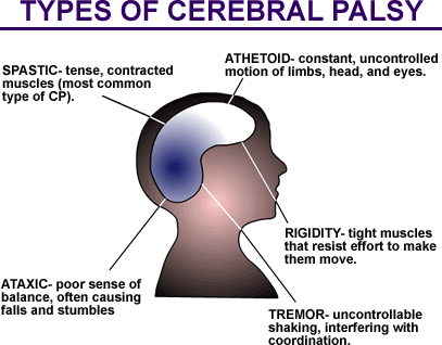
When a child is about 2 years old, the doctor may be able to diagnose the type of CP he or she has. There are four main types:


• Spastic:
In spastic CP, the most common form, there is increased muscle tone/tightness. Legs, arms and back are stiff and contracted, making movement difficult. If both legs are affected (spastic diplegia), tight muscles in the hips and legs cause legs to turn inward and cross at the knees when walking. If only one side of the body is affected (spastic hemiplegia), the arm is often more severely affected than the leg. If all four limbs and the trunk are affected (spastic quadriplegia or total body involved), the muscles of the mouth and tongue may also be affected.


• Athetoid: 
(dyskinetic): In athetoid CP there is low muscle tone/looseness, leading to uncontrolled movements of the entire body. It may be difficult to sit straight or walk.


• Ataxic: 
A rare form, ataxic CP affects balance and depth perception. There is poor coordination, unsteady walking and difficulty with precise movements such as using a pen or buttoning a shirt.


• Mixed: 
In mixed CP, there are symptoms of both spastic and athetoid CP. Some muscles are tight and others are loose. There is both stiffness and involuntary movements.


Treatment 


Sometimes people with cerebral palsy need help from their families throughout their lives. If your child has CP, you may want to work with a treatment team of health care professionals to identify and meet the child's individual needs. The goal is to help your child reach his or her potential and maximize independence. Treatment teams often include:

• Orthopaedic surgeons to treat problems with bones, muscles, tendons, nerves    or joints.
• Physical therapists to improve movement and strength.
• Occupational therapists to teach skills required for daily living (i.e., eating and    dressing).
• Speech and language specialists to treat communication problems.
• Social workers to help locate community assistance and education programs.
• Psychologists to help patients and families cope with stress and/or behavioral    therapists to foster social and emotional development.
• Other medical specialists such as eye doctors, neurologists and nutritionists.


Nonsurgical Treatment 


• Physical therapy. 
Physical therapy (PT) may begin immediately after diagnosis to help your child learn skills like sitting, walking or using a wheelchair. It may also help improve muscle strength, balance and coordination; and prevent muscles from shortening. Stretching muscles may help prevent contracture. Sometimes PT may involve activities like swimming and horseback riding to tone muscles.

• Braces, splints, and casts. 
Using braces, splints or casts may improve joint mobility and stability, prevent
contracture and improve hand or leg function. Braces can compensate for muscle imbalance.

• Botox.
This drug can be injected into spastic muscles to loosen them up. It is especially
helpful in children younger than 5 and when used together with casting.

• Medications.
Medications may control or prevent seizures or muscle spasms, ease muscle stiffness or reduce abnormal movements.

• Mechanical aids. 
Special machines like computerized communication devices or simple aids like
velcro-fastened shoes or crutches may help a child overcome impairments.


Coping Strategies 


Parents of children with cerebral palsy or other disabilities often feel guilt and/or grief about the
child's condition. Some tips to help parents cope:

• Learn all you can about CP. You may want to join a family support group or get    help from professionals. Stay informed about new treatments and    technologies.
• Work with professionals at your child's school to develop an individualized       plan that meets his or her needs and abilities.
• Love and encourage your child. Family support and personal determination are
   important factors in achieving long term goals. Treat your child like a normal    kid. Go places and have fun. Be patient, and keep hoping for improvement.
• Get help from family and friends. Caring for a child with CP is hard work. Teach
   others how to do it so you can take breaks.


Surgical Treatment 


If contractures are severe, a child with CP may need surgery to lengthen affected muscles.
Surgery may also help if tightly contracted muscles cause stress to joints and lead to deformities or dislocations. Some children with CP need surgery to correctly position their arms or legs, or to correct curvature of the spine. Other surgical options for spasticity management include selective dorsal rhizotomy and intrathecal baclofen pumps. These procedures are usually done by neurosurgeons.

What ethical, regulatory, and security challenges accompany AI implementation in healthcare settings?

 What ethical, regulatory, and security challenges accompany AI implementation in healthcare settings? The implementation of AI in healthcar...